How does coronavirus leave the cell?
Freiburg, Sep 11, 2020
Freiburg pharmacologist Prof. Dr. Robert Grosse studies how the SARS-CoV-2 virus changes structures inside cells, and is contributing to major international studies. He is mainly focusing on a specific enzyme that interacts with coronavirus. Grosse is a professor at the Institute of Experimental and Clinical Pharmacology and Toxicology and also undertakes research at the Freiburg Cluster of Excellence CIBSS – Centre for Integrative Biological Signalling Studies.
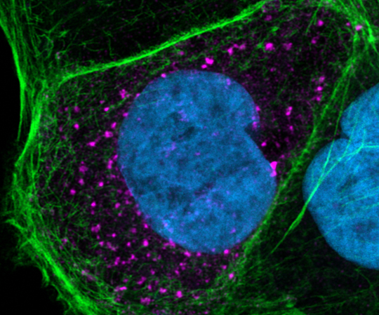
The pink dots are molecules of the SARS-CoV-2 virus, within a human cell. Robert Grosse and his team are studying how these molecules affect the cell skeleton. Source: Robert Grosse
The photograph shows a human intestinal epithelial cell against a very black background. In the interior of the cell are pink dots, and there are small protrusions at its edge. It looks a little as if an invisible force is pulling the cell membrane outwards. Robert Grosse and his team made the photograph with a laser scanning microscope more than four months ago now. These seemingly harmless dots in the picture are molecules of the new coronavirus, Grosse explains. With the photograph he wanted to discover what SARS-CoV-2 does to the cell skeleton, that is the structure that keeps cells stable but also mobile.
Grosse has been researching the protein actin, which forms an important part of the cell skeleton, for years. Occasionally he has also researched how viruses, specifically HIV and Vaccinia, change the cell skeleton. With SARS-CoV-2 Grosse suddenly had new material for his research. Together with his PhD student Svenja Ulferts he got to work in March 2020. With his connections in virology, Grosse and Ulferts were also able to use the S3 laboratory (S3 stands for security level 3) at the Freiburg University Medical Center. They were supported in this task by Sebastian Weigang, who is working on his doctorate there. The team had barely taken its first microscopic photos before they became part of an international research association, working on a major study. It was initiated by Pedro Beltrao of the European Institute of Bioinformatics at the University of Cambridge, and by Nevan Krogan who heads the Quantitative Biosciences Institute in San Francisco.
Fine lines branching like fingers
This study also focuses on SARS-CoV-2 and the protein casein kinase 2 (CK2) which has a fascinating interaction with the virus. Fascinating, because the virus has a strikingly powerful activating effect on the enzyme within cells. The Freiburg team has contributed microscopic analyses documenting precisely this interaction to the study. They show fine lines leading from the cell and branching at the tip like fingers. Grosse also calls these lines roads: “The virus changes the actin structure of the cell, pulls it apart at one point or another.” Although it looks as if these roads transport the virus and CK2 out of the cell, the function of these finger structures is still unclear. So far they have only been able to see that CK2 is contained in these structures and co-localized with the virus proteins. That means, there is a biochemical compound.
Grosse’s finding corresponds with the photographs taken by American researcher Elisabeth Fisher, who took very similar images at the same time – however she used an electron microscope. “In these high resolution images it looks as if the virus is actually leaving the cell,” says Grosse, immediately adding, “At the moment this is nothing more than a hunch, after all we are working with ‘snapshots’ and not film which could document progression.”
The right inhibitor is crucial
The interaction of cell, virus and CK2 is also fascinating because it could give rise to potential treatments for covid-19. This would be the main achievement of the study, Grosse says. A first step is to examine specifically which kinases are activated in the cell that is infected with SARS-CoV-2, in order to list the substances that inhibit these kinases. Some of these pharmacological inhibitors have already been tested in clinical trials, however not in connection with covid-19, but mainly for tumor therapies. One such substance is silmitasertib which inhibits CK2, and Grosse will be investigating it in the coming months. If it in fact turns out that CK2 is responsible for the formation of the branched roads and the virus is also passed on via these roads to neighboring cells, this process could potentially be stopped with the right inhibitors.
However this is all still very speculative, Robert Grosse stresses. Nevertheless research is continuing in this direction. Or more accurately in several directions: in order to keep CK2 as small as possible, Grosse and his team are currently testing several methods. In addition the researchers want to inhibit other enzymes activated by the virus that polymerize the actin fibers in the cell skeleton, that is pull them apart. As regards SARS-CoV-2, Grosse will continue to collaborate with other scientists in future. Including with Beltrao: they want to dig deeper, to understand what the virus is doing to the cell. Grosse is delighted about this cooperation and has found the international connection extremely exciting. Working on research in such a large team across many national boundaries has been fantastic, and has produced an incredible vitality.
Stephanie Streif