The virtual microscope
Freiburg, Aug 11, 2021
Stained tissue samples help doctors make more precise diagnoses for their patients. With the aid of a microscope, structural changes in tissue can be detected. In medicine, this field is called histology and is an important part of medical studies. Together with Ulm University, Prof. Dr. Andreas Vlachos and Dr. Bernd Heimrich from the Faculty of Medicine are developing an app for students that helps them learn to recognize and analyze tissue abnormalities.
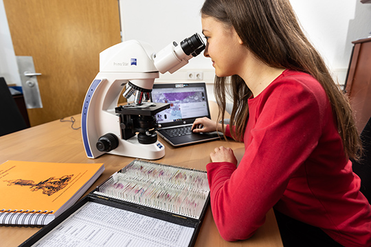 Medical student Phyllis Stöhr is part of the team that entered the Freiburg data sets into the app. Photo: Jürgen Gocke
Prof. Dr. Andreas Vlachos lists “the clear view towards the digital” among the few helpful improvements that has accompanied Corona. The head of the Department of Neuroanatomy at the Institute of Anatomy and Cell Biology at the University of Freiburg thinks that good things have been initiated. But the pandemic is not responsible for all digitalization projects. The software program “MyMi.mobile,” for example, a cooperation between Ulm University and the University of Freiburg, was created independently. It is an app with access to a database in which histological samples, i.e. tissue samples, are available in digital form. Students learning how the human body is constructed can now access this app. The latest version of which was released in July 2020. It is available to them anytime and anywhere.
“The first developmental steps date back ten years,” says Dr. Bernd Heimrich from the Department of Neuroanatomy. He is responsible for coordinating instruction. “Prof. Dr. Stefan Britsch initiated it in Ulm at the time,” he says. The cooperation came about because Britsch and Heimrich knew each other well from the Charité Universitätsmedizin Berlin. “It’s incredible the foresight Stefan Britsch showed back then,” adds Vlachos: “The program was structured to be modular and scalable.”
Preserving teaching culture
Modular means that new building blocks can be added in an uncomplicated way, for example the histological samples from Freiburg. They were not integrated into the Ulm inventory, but added as independent data sets. “Each institute has its own identity and teaching culture,” explains Vlachos. The fact that the app allows such cultures to stand side by side instead of unifying them is important, he says. It facilitates future collaborations, he says. This, in turn, he says, leads to an increase in data sets and possible uses, making the app even more useful. In the future, artificial intelligence systems will also help students learn. “Talks are underway with other cooperation partners at universities in Baden-Württemberg,” says Heimrich. “After all, such mergers also save money,” adds Vlachos. “That way we aren’t having to reinvent the wheel ten times over.”
 With the "MyMi.Mobile" app, students can consolidate their knowledge of histology from home. Photo: Jürgen Gocke
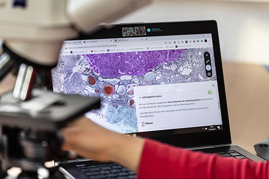
The team that entered the data sets in Freiburg includes Phyllis Stöhr, a student of human medicine. She got to know the app in her second semester and was immediately convinced of its value for teaching. “That’s why I attended a training course on data entry in Ulm during the summer vacations, so that I could help shape the further development of MyMi.mobile from the third semester onwards.” Medical students examine different tissue samples during their training. “During classroom instruction, everyone sits at the microscope and receives instructions from the lecturers.” In MyMi.mobile, these notes are replaced by “annotations” that Stöhr, among others, created. “We marked important structures, and there’s also accompanying text.” Clicking on a small owl symbol gives access to more in-depth information.
The app has a virtual high-resolution microscope with a continuous zoom function and offers students other features such as “Find the structure” for training visual skills and “Make the diagnosis.” No other app in the German-speaking world does anything like this.
Making reliable diagnoses
Phyllis Stöhr now works with MyMi.mobile herself and knows the feedback from fellow students. "It's very good, the app has helped many, especially when classroom instruction was not possible." Bernd Heimrich can back up this assessment with data. “Last year, a total of just under 1,000 students at both universities accessed MyMi.mobile more than 200,000 times. They use the app regularly and over and over again.” But Stöhr, a medical student, does not believe that the app will one day completely replace classroom instruction. She agrees with Andreas Vlachos that the digital has limits. “Only manual work at the microscope and scientifically guided independent analysis without clues and markers deepens histological knowledge in such a way that you can later make and understand reliable diagnoses.”
Mathias Heybrock