Silent Yet Eloquent Models
Freiburg, Feb 07, 2018
Photo: Jürgen Gocke
A shoulder, part of a back, half a leg. You might assume that this a description of a slaughterhouse – but you’d be mistaken. These objects are on display in a seminar room at the Department of Dermatology of the Freiburg University Medical Center.
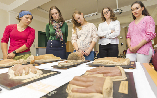
Like doctors on their rounds: Medical students study moulages as case studies depicting the typical symptoms of a disease. Photo: Jürgen Gocke
Dermatologist Dr. Martin Faber has taken the objects out of a metal cabinet and placed them carefully on a table. They are moulages, realistic wax or plastic molds of human body parts that used to serve as models for training medical students. In the past years these molds have been finding their way back into the classroom. Faber asks the students to describe and compare the symptoms of various skin diseases through examination of selected moulages. The course, which is coordinated by adjunct faculty member Prof. Dr. Christoph Schempp, received the teaching award of the University of Freiburg’s Faculty of Medicine for outstanding instruction in medicine in 2017.
Faber has been offering moulage courses as part of a compact practical course in dermatology for the past ten years. The students showed interest in the courses from the start, he says, because “at some point they want to see something tangible. It’s one thing to see a photograph but a completely different thing to stand before a limb or a body part molded in wax.” One views a photograph or a PowerPoint presentation passively, as if in a movie theater, but it is possible simply by shifting one’s position to develop a physical relationship with a three-dimensional object. “One learns, for instance, that an object looks different depending on whether it is observed in natural or artificial light.”
Small Groups
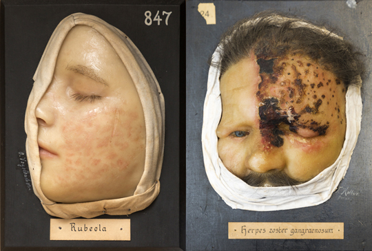
“As a person who is given to contemplation, I recommend studying things in peace as a source of pleasure. I discover an entire cosmos when processing visual impressions.” Martin Faber also appreciates the artistic aspects of moulages. The important thing is the colors. The objects should be painted to look realistic. In addition, the cloth draped around the head of a child in a moulage depicting measles reminds him of the portrait of a saint. It is perhaps no accident that the objects are signed like paintings.
Faber teaches the graded practical course in small groups. Two of a total of 30 contact hours are reserved exclusively for examining moulages. He plans to continue teaching the moulage course for two more years before going into retirement. However, the moulages have already been integrated into the “case-based learning” approach practiced by the head physicians at the department, ensuring the continued existence of this special form of teaching in the future. Faber is grateful that the University Medical Center agreed to cover the high costs of cleaning and restoring the moulages, and he is absolutely certain that the students still see the good sense and use in training their skills with these silent yet at the same time eloquent models of skin diseases.
Unlike bedside teaching with real patients, moulages provide educational examples that are available at all times. Photos: Jürgen Gocke
The course also includes an interactive e-learning program using photographs of the moulages as case studies that is available to the students on the learning platform ILIAS. The program shows and explains the efflorescences, the smallest macroscopic unit used in dermatological descriptions. The students then take a quiz to test whether they have learned the specialist terminology they have already been introduced to while examining the moulages in the course. The e-learning program with moulages therefore serves as a means of reviewing, extending, and testing the skills practiced in the moulage course.
Lifelike and true-to-scale models
The use of moulages in medical instruction was increasingly regarded as obsolete in the decades following the Second World War. In Freiburg the wax models were put in storage in the 1990s. Other universities stopped using them even earlier. The morphological teaching model in dermatology began going out of fashion in the mid 20th century and was replaced increasingly by functional approaches, and later by the molecular biological model. Technological advances such as high-quality color photography also contributed to the decline in the use of moulages. In Freiburg this development was compounded by a lack of space due to the expansion of the Allergy Department. Today a large part of the 832 moulages owned by the University Medical Center are stored in the cellar. Selected specimens are kept in the metal cabinet mentioned above, and others are on display at the University of Freiburg’s Uniseum.
The first signs of a renaissance in the use of moulage for medical instruction in Germany appeared two decades ago. The reasons for the second career these usually antique objects are now enjoying are immediately apparent: Christoph Schempp values moulages not just as “lifelike and true-to-scale models of skin diseases with great educational value” but also because they can serve as a “realistic and graphic illustration of even rare diseases.” Do these advantages justify the high costs of restoration? “Definitely,” says Schempp. “The skin is something that is readily available for close examination. The only thing better than moulages is real patients. They can’t be topped by books or electronic media.”
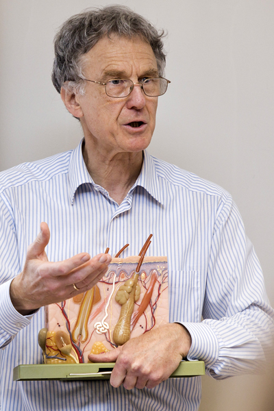
Martin Faber has been offering moulage courses as part of a compact practical course in dermatology for the past ten years. The students showed interest in the courses from the start. Photo: Jürgen Gocke
Valuable cultural treasure
The starting point for the revival of moulage was a 1995 study by Prof. Dr. Thomas Schnalke, director of the Institute for the History of Medicine at the Charité Teaching Hospital in Berlin. The study is now regarded as a standard work on the history of moulages. Doctors at Charité also initiated a “moulage working group” several years ago. Its members now include ten dermatology departments that are again using moulages in the classroom. The reason for the great number of 832 moulages in Freiburg is that Prof. Dr. Eduard Jacobi, then head of the Department of Dermatology, published the first atlas of skin diseases in 1906 using color photographs. The exposure times for color photography were so long around the turn of the 20th century that it was impossible to take sharp pictures of patients.
To make high-quality color reproductions in his skin atlas, Jacobi therefore had numerous moulages made and used them as immobile models for his photographs. Many of the Freiburg moulages are of excellent quality – no doubt in large part due to the fact that Freiburg’s first moulage maker was an academically trained artist. Schempp sees the objects as a “valuable cultural treasure of high artistic and educational value.” He also holds the work of Martin Faber in high regard: “With his great devotion to Freiburg’s moulages, Martin Faber has rendered a great service to the department. We owe it largely to his initiative that our moulage collection has been restored to its old glory. In this way, we were able to integrate our moulages into practical instruction and e-learning.”
Hans-Dieter Fronz